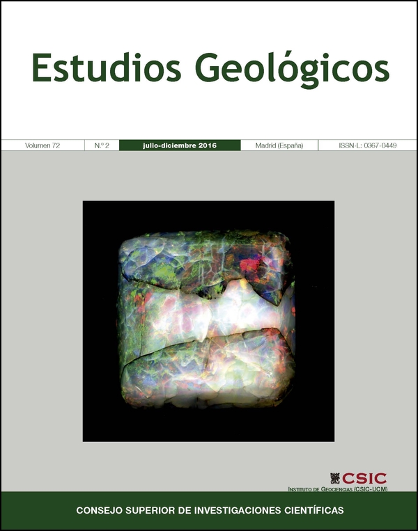
Solving geological and historical puzzles with advanced gemologic techniques: The Franco Dávila (1772) precious opal case
DOI:
https://doi.org/10.3989/egeol.42459.410Keywords:
opal, gemology, historic collections, Non-destructive techniquesAbstract
The large precious opal weighting 33 grams fitted in a silver jewel and exposed to visitors at the Museo Nacional de Ciencias Naturales (MNCN) is well documented in: (i) its own mounting (1772), (ii) at the 775 document of the Archive of the MNCN and (iii) the 395 specimen described in the of Pedro Franco Dávila catalogue. The X-ray diffractogram (XRD) performed onto the opal block is very similar to other opals of volcanic origin containing varied amounts of cristobalite, tridymite and amorphous silica. The Raman spectrum shows a band peaked at 242, 343 and 416 cm-1 associated with O-Si-O stretching groups; other spectral band peaked at 780 and 819 cm-1 corresponding to vibration of symmetrical O-Si-O rings of 3 and 4 link members, plus other minor bands. The Raman spectrum is also very similar to those observed in Mexican opals of volcanic origin containing an spectral band of stretching nodes v1 (OH) at 3233, 3393, 3511, 3628 cm-1 related to OH groups with hydrogen bonds of isolated silanol groups. The interferometric confocal dual microscope 3D (MCI3D), which is a nondestructive facility of high resolution and LED technology reveals the geometry of graver tools on the silver jewel and the computed tomography X-ray highlights the opal cutting as a squared princess type and silver chloride infillings of a crack probably caused by a shock on a corner. Under the scanning electron microscope we observed barite, sealed veins of silica rich in Mn and opal with high contents of Al and K which, along with the historical data, the piece can be attributed to the historical site of opals hosted in Slovakia andesite rocks, this data explains the optical light behavior in the cabochon. The silver jewel has large amounts of Hg and AgCl indicating amalgam method. In addition the natural AgS2 phases probably come from Nueva España (year 1772) in full production of silver in such time. The association of new analytical non-destructive techniques combines the preservation of samples together with significant analytical data allowing us to deduce genetic mineral processes, provenances and manufacturing techniques of materials. These facilities allow the characterization, interpretation, conservation and enhancement of cultural and archaeological heritage.
Downloads
References
Alonso, P.J.; Halliburton, L.E.; Kohnke, E.E. & Bossoli, R.B. (1983). X-ray-induced luminescence in crystalline SiO2. Journal of Applied Physics, 54: 5369–5375. https://doi.org/10.1063/1.332715
Artioli, G. & Quartieri, S. (2016). The Contribution of Geoscience to Cultural Heritage Studies. Elements, 12 (1): 13–18. https://doi.org/10.2113/gselements.12.1.13
Auer, B.M. & Skinner, J.L. (2008). IR and Raman spectra of liquid water: theory and interpretation. Journal of Chemical Physics, 128 (22): 224511. https://doi.org/10.1063/1.2925258 PMid:18554033
Belem, T.; Homand-Etienne F. & Souley M. (2000). Quantitative parameters for rock joint surface roughness. Rock Mechanics and Rock Engineering, 33 (4): 217–242. https://doi.org/10.1007/s006030070001
Brewster, S.D. (1845). An account of the cause of the colors in precious opal. Journal of the Franklin Institute, 40 (3): 195. https://doi.org/10.1016/0016-0032(45)90571-0
Cagnetti, A. (2009). Experimental survey on fluid brazing in ancient goldsmith's art. International Journal of Materials Research, 100 (1): 81–85. https://doi.org/10.3139/146.101783
Calatayud-Arinero, M.A. (1987). Catálogo de Documentos del Real Gabinete de Historia Natural (1752–1786). CSIC, Madrid, p. 277.
Canet, C.; Camprubí, A.; González-Partida, E.; Linares, C.; Alfonso, P.; Pi-eiro-Fernández, F. & Prol-Ledesma, R.M. (2009). Mineral assemblages of the Francisco I. Madero Zn-Cu-Pb-(Ag) deposit, Zacatecas, Mexico: implications for ore deposit genesis. Ore Geology Reviews, 35 (3–4): 423–435. https://doi.org/10.1016/j.oregeorev.2009.02.004
Carey, D.M. (1998). Measurement of the Raman spectrum of liquid water. Journal of Chemical Physics, 108: 2669–2675. https://doi.org/10.1063/1.475659
Caucia, F.; Ghisoli, C.; Marinoni, L. & Bordoni, V. (2013). Opal, a beautiful gem between myth and reality. Neues Jahrbuch fur Mineralogie, Abhandlungen, 190 (1): 1–9. https://doi.org/10.1127/0077-7757/2012/0226
Caucia, F.; Marinoni, L.; Leone, A. & Adamo, I. (2013). Investigation on the gemological, physical and compositional properties of some opals from Slovakia (Hungarian opals). Periodico di Mineralogia, 82 (2): 251–261.
Crettenden, P.P.; Flintoft, M.W.; Ewen, S.J. & Watkins, D.C. (1979). The opal industry in South Australia. Mineral Resources Review (Department of Mines, South Australia), 151: 18–29.
Chutas, N.I. & Sack, R.O. (2004). Ore genesis at La Colorada Ag-Zn-Pb deposit in Zacatecas, Mexico. Mineralogical Magazine, 68 (6): 923–937. https://doi.org/10.1180/0026461046860231
Darragh, P.J.; Gaskin, A.J.; Terrell, B.C. & Sanders, J.V. (1966). Origin of precious opal. Nature, 209 (5018): 13–16. https://doi.org/10.1038/209013a0
Demortier, G.; Fernández-Gómez, F.; Ontalba Salamanca, M.A. & Coquay, P. (1999). PIXE in an external microbeam arrangement for the study of finely decorated tartesic gold jewellery items. Nuclear Instruments and Methods in Physics Research, Section B: Beam Interactions with Materials and Atoms, 158 (1–4): 275–280. https://doi.org/10.1016/S0168-583X(99)00311-0
Dobado, R. & Marrero G.A. (2011). The role of the Spanish imperial state in the mining-led growth of Bourbon Mexico's economy. Economic History Review, 64 (3): 855–884. https://doi.org/10.1111/j.1468-0289.2010.00555.x
Fisher, J.R. (1977). Silver mines and silver miners in colonial Peru, 1776–1824. Centre for Latin American Studies, University of Liverpool, 150 p.
Franco Davila, P. (1784). A monsieur Boytet Consul General de Francia en esta corte, actualmente en Paris. Madrid a 7 de agosto de 1784. Copiador de Cartas. Legajo nº 18, página 1. Archivo del Museo Nacional de Ciencias Naturales. Madrid.
García-Guinea, J.; Fernández-Cortés, A.; Álvarez-Gallego, M.; García-Antón, E.; Casas-Ruiz, M.; Blázquez-Pérez, D.; Teijón, O.; Cuezva, S.; Correcher, V. & Sánchez-Moral, S. (2013). Leaching of uranyl-silica complexes from the host metapelite rock favoring high radon activity of subsoil air: Case of Casta-ar cave (Spain). Journal of Radioanalytical and Nuclear Chemistry, 298 (3): 1567–1585. https://doi.org/10.1007/s10967-013-2587-7
Gorton, N.T.; Walker, G. & Burley, S.D. (1997). Experimental analysis of the composite blue cathodoluminescence emission in quartz. Journal of Luminescence, 72–74: 669–671. https://doi.org/10.1016/S0022-2313(96)00242-6
Götze, J.; Plötze, M. & Habermann, D. (2001). Origin, spectral characteristics and practical applications of the cathodoluminescence (CL) of quartz - a review. Mineralogy and Petrology, 71 (3): 225–250. https://doi.org/10.1007/s007100170040
Hachisu, S. & Yoshimura, S. (1980). Optical demonstration of crystalline superstructures in binary mixtures of latex globules. Nature, 283 (5743): 188–189. https://doi.org/10.1038/283188a0
Hsu, T.; Lucas, A. & Pardieu, V. (2015). Splendor in the outback: A visit to Australia's opal fields. Gems & Gemology, 51 (4): 418–427.
Ilieva, A.; Mihailova, B.; Tsintsov, Z. & Petrov, O. (2007). Structural state of microcrystalline opals: A Raman spectroscopic study. American Mineralogist, 92 (8–9): 1325–1333 . https://doi.org/10.2138/am.2007.2482
Itoh, C.; Tanimura, K. & Itoh, N. (1988). Optical studies of self-trapped excitons in SiO2. Journal of Physics C: Solid State Physics, 21 (26): 4693–4702. https://doi.org/10.1088/0022-3719/21/26/017
Jones, C.E. & Embree, D. (1976). Correlations of the 4.77–4.28-eV luminescence band in silicon dioxide with the oxygen vacancy. Journal of Applied Physics, 47: 5365–5371. https://doi.org/10.1063/1.322562
Jones, J.B.; Sanders, J.V. & Segnit, E.R. (1964). Structure of opal. Nature, 204 (4962): 990–991. https://doi.org/10.1038/204990a0
Jones, J.B. & Segnit, E.R. (1971). The nature of opal I. nomenclature and constituent phases. Journal of the Geological Society of Australia, 18 (1): 57–67. https://doi.org/10.1080/00167617108728743
Kalceff, M.A.S. & Phillips, M.R. (1995). Cathodoluminescence microcharacterization of the defect structure of quartz. Physical Review B, 52 (5): 3122–3134. https://doi.org/10.1103/PhysRevB.52.3122
Ketcham, R.A. & Carlson, W.D. (2001). Acquisition, optimization and interpretation of x-ray computed tomographic imagery: Applications to the geosciences. Computers & Geosciences, 27 (4): 381–400. https://doi.org/10.1016/S0098-3004(00)00116-3
Kita, N.T.; Ushikubo, T.; Fu, B. & Valley, J.W. (2009). High precision SIMS oxygen isotope analysis and the effect of sample topography. Chemical Geology, 264 (1–4): 43–57. https://doi.org/10.1016/j.chemgeo.2009.02.012
Krbetschek, M.R.; Götze, J.; Dietrich, A. & Trautmann, T. (1997). Spectral information from minerals relevant for luminescence dating. Radiation Measurements, 27 (5–6): 695–748. https://doi.org/10.1016/S1350-4487(97)00223-0
Langer, K. & Flörke O.W. (1974). Near infrared absorption spectra (4000-9000 cm-1) of opals and the role of water in these SiO2.nH2O minerals. Fortschritte der Mineralogie, 52: 17–51.
Lee, H.S.; Park, Y.J.; Cho, T.F. & You, K.H. (2001). Influence of asperity degradation on the mechanical behavior of rough rock joints under cyclic shear loading. International Journal of Rock Mechanics & Mining Sciences, 38 (7): 967–980. https://doi.org/10.1016/S1365-1609(01)00060-0
Luff, B.J. & Townsend, P.D. (1990). Cathodoluminescence of synthetic quartz. Journal of Physics: Condensed Matter, 2: 8089–8097. https://doi.org/10.1088/0953-8984/2/40/009
Macdonald, R.M. (1904). The opal formations of Australia. Scottish Geographical Magazine, 20 (5): 253–261. https://doi.org/10.1080/14702540408554624
Mango, H.; Arehart, G.; Oreskes, N. & Zantop, H. (2014). Origin of epithermal Ag-Au-Cu-Pb-Zn mineralization in Guanajuato, Mexico. Mineralium Deposita, 49 (1): 119–143. https://doi.org/10.1007/s00126-013-0478-z
Martini, M.; Paleari, A.; Spinolo, G. & Vedda, A. (1995). Role of [AlO4]0 centers in the 380-nm thermoluminescence of quartz. Physical Review B, 52 (1): 138–142. https://doi.org/10.1103/PhysRevB.52.138
Meixner, A.J. (2016). The Nobel Prize in Chemistry 2014 for the development of super-resolved fluorescence microscopy. Analytical & Bioanalytical Chemistry, 407 (7): 1797–1800. https://doi.org/10.1007/s00216-014-8444-x PMid:25633213
Moncada, D.; Mutchler, S.; Nieto, A.; Reynolds, T.J.; Rimstidt, J.D. & Bodnar, R.J. (2012). Mineral textures and fluid inclusion petrography of the epithermal Ag-Au deposits at Guanajuato, Mexico: Application to exploration. Journal of Geochemical Exploration, 114: 20–35. https://doi.org/10.1016/j.gexplo.2011.12.001
Murali, K.V.R.M.; Naik, V.B. & Datta, D. (2015). Gallium-nitride-based light-emitting diodes: 2014 Nobel Prize in Physics. Resonance, 20 (7): 605–616. https://doi.org/10.1007/s12045-015-0219-y
Ostrooumov, M.; Fritsch, E.; Lasnier, B. & Lefrant, S. (1999). Raman spectroscopy of opals: diagnostics and classification aids. European Journal of Mineralogy, 11 (5): 899–908. https://doi.org/10.1127/ejm/11/5/0899
Ponzio de Léon, C.A. (1998). Interpretación económica del último periodo colonial Mexicano. Trimestre Economico, 65 (257): 99–125.
Ramos-Arroyo, Y.R.; Prol-Ledesma, R.M. & Siebe-Grabach, C. (2004). Características geológicas y mineralógicas e historia de extracción del Distrito de Guanajuato, México. Posibles escenarios geoquímicos para los residuos mineros. Revista Mexicana de Ciencias Geologicas, 21 (2): 268–284.
Rau, R.C. & Amaral, E.J. (1969). Electron microscopy of precious opal. Metallography, 2 (4): 323–328. https://doi.org/10.1016/0026-0800(69)90062-7
Remond, G.; Cesbron, F.; Chapoulie, R.; Ohnenstetter, D.; Roques-Carmes, C. & Schvoerer, M. (1992). Cathodoluminescence applied to the microcharacterization of mineral materials: A present status in experimentation and interpretation. Scanning Microscopy, 6: 23–68.
Rink, W.J.; Rendell, H.; Marseglia, E.A.; Luff, B.J. & Townsend, P.D. (1993). Thermoluminescence spectra of igneous quartz and hydrothermal vein quartz. Physics & Chemistry of Minerals, 20 (5): 353–361. https://doi.org/10.1007/BF00215106
Rondeau, B.; Fritsch, E.; Guiraud, M. & Renac, C. (2004). Opals from Slovakia (Hungarian opals): A re-assessment of the conditions of formation. European Journal of Mineralogy, 16: 789–799. https://doi.org/10.1127/0935-1221/2004/0016-0789
Sanders, J.V. (1964). Colour of precious opal. Nature, 204 (4964): 1151–1153. https://doi.org/10.1038/2041151a0
Sanders, J.V. & Murray, M.J. (1978). Ordered arrangements of spheres of two different sizes in opal. Nature, 275 (5677): 201–203. https://doi.org/10.1038/275201a0
Sigel Jr, G.H. & Marrone, M.J. (1981). Photoluminescence in as-drawn and irradiated silica optical fibers: an assessment of the role of non-bridging oxygen defect centers. Journal of Non-Crystalline Solids, 45 (2): 235–247. https://doi.org/10.1016/0022-3093(81)90190-3
Smallwood, A.G.; Thomas, P.S. & Ray, A.S. (1997). Characterisation of sedimentary opals by Fourier transform Raman spectroscopy. Spectrochimica Acta - Part A: Molecular & Biomolecular Spectroscopy, 53 (13): 2341–2345. https://doi.org/10.1016/S1386-1425(97)00174-1
Spooner, N.A. & Questiaux, D.G. (2000). Kinetics of red, blue and UV thermoluminescence and optically-stimulated luminescence from quartz. Radiation Measurements, 32 (5–6): 659–666. https://doi.org/10.1016/S1350-4487(00)00067-6
Stevens-Kalceff, M.A. (2009). Cathodoluminescence microcharacterization of point defects in ?-quartz. Mineralogical Magazine, 73 (4): 585–605. https://doi.org/10.1180/minmag.2009.073.4.585
Stevens-Kalceff, M.A. (2013). Cathodoluminescence microanalysis of silica and amorphized quartz. Mineralogy and Petrology, 107 (3): 455–469. https://doi.org/10.1007/s00710-013-0275-5
Sun, Q. (2009). The Raman OH stretching bands of liquid water. Vibrational Spectroscopy, 51 (2): 213–217. https://doi.org/10.1016/j.vibspec.2009.05.002
Tamla, Ü. & Varkki, H. (2009). Learning the technologies of making beaded wire. Estonian Journal of Archaeology, 13 (1): 36–52. https://doi.org/10.3176/arch.2009.1.03
Walrafen, G.E.; Fisher, M.R.; Hokmabadi, M.S. & Yang, W.H. (1986). Temperature dependence of the low- and high-frequency Raman scattering from liquid water. The Journal of Chemical Physics, 85: 6970–6982. https://doi.org/10.1063/1.451384
Wellington, S.L. & Vinegar, H.J. (1987). X-Ray Computerized Tomography. Journal of Petroleum Technology, 39 (8): 885–898. https://doi.org/10.2118/16983-PA
Published
How to Cite
Issue
Section
License
Copyright (c) 2016 Consejo Superior de Investigaciones Científicas (CSIC)

This work is licensed under a Creative Commons Attribution 4.0 International License.
© CSIC. Manuscripts published in both the printed and online versions of this Journal are the property of Consejo Superior de Investigaciones Científicas, and quoting this source is a requirement for any partial or full reproduction.All contents of this electronic edition, except where otherwise noted, are distributed under a “Creative Commons Attribution 4.0 International” (CC BY 4.0) License. You may read here the basic information and the legal text of the license. The indication of the CC BY 4.0 License must be expressly stated in this way when necessary.
Self-archiving in repositories, personal webpages or similar, of any version other than the published by the Editor, is not allowed.